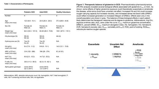
OralL-glutamine (Endari®) has been approved by the US-FDA to reduce the acute complications of sickle cell disease (SCD) in patients at least 5 years of age. L-glutamine has several roles in the body including de novo synthesis of intracellular glutathione which acts as an antioxidant. However, the mechanisms of action by which L-glutamine could reduce complications in SCD require further investigation. Additionally, there are no known biomarkers to assess response to L-glutamine therapy.
We conducted an open-label, dose-ascending trial (NCT04684381) of L-glutamine in pediatric (n=4) and adult (n=4) participants with SCD as well as in adult healthy volunteers (n=4) (Table 1). Over a three-week trial period with 4 study visits, serial blood samples were collected to define the pharmacokinetics (PK), pharmacodynamics (PD), and PK-PD interactions of L-glutamine using a broad panel of laboratory investigations including amino acid concentrations, blood counts, percentage of dense red blood cells (%DRBC), whole blood viscosity, osmotic and oxygen gradient ektacytometry, and reactive oxygen species (ROS) of reticulocytes (ROS retic) and mature red blood cells (ROS RBC).
In PK-PD analysis of amino acids, we focused analysis on arginine (Arg), citrulline (Cit), and ornithine (Orn) based on the hypothesis that L-glutamine improves endothelial function by increasing arginine bioavailability and augmenting nitric oxide (NO) production. Peak arginine concentration was directly correlated with both the maximum L-glutamine concentration (C max p=0.001) and area-under-the curve (AUC) (p=0.047) indicating improvement in arginine bioavailability, but there was no significant linear correlation with either the Arg:Orn or Arg:(Orn+Cit) ratios.
In PK-PD analysis of osmotic gradient ektacytometry, L-glutamine C max was directly correlated with the elongation maximum (EI max), noted in all participants at visit 4 (p=0.033). In SCD participants, L-glutamine C max was inversely correlated with O hyper (osmolality at EI max/2) at visits 3 (p=0.044) and 4 (p=0.004). In PK-PD analysis of oxygen gradient ektacytometry in SCD participants, L-glutamine C max was inversely correlated with the point of sickling (POS) at visit 3 (p=0.005) but not at visit 4 (p=0.054). Together, these findings suggest that although L-glutamine may decrease cellular hydration (decreased O hyper) it may increase RBC deformability (increased EI max) and delay the onset of sickling after hypoxia (decreased POS).
In PK-PD analysis of viscometry, higher L-glutamine C max was associated with higher whole blood viscosity measurements at all shear rates (p<0.05). However, L-glutamine C max was also directly correlated with hematocrit-to-viscosity ratio (HVR) at the highest shear rates in the overall population at visit 4 (p<0.05). In addition, L-glutamine C max for SCD participants was associated with increased hemoglobin concentration, an effect that was observed at visit 4 (p=0.025), and increased %DRBC, an effect detected at visits 3 (p=0.008) and 4 (p=0.007).
In PK-PD analysis of ROS, L-glutamine AUC was inversely correlated with ROS Retic (p=0.024), but not ROS RBC, in all participants at visit 3. When considered as total ROS Retic (calculated as ROS retic × absolute reticulocyte count), there was a similar inverse correlation with glutamine AUC in SCD participants, although not statistically significant. In contrast to all other biomarkers, L-glutamine C max was not correlated with any ROS measurement.
This PK-PD analysis of L-glutamine reveals biological effects that alter RBC characteristics and could modify SCD-related complications, with a complex interplay summarized in Figure 1. Prospective studies can validate these biomarkers and be used to monitor the effects of L-glutamine therapy in SCD patients.
Disclosures
Kalfa:Forma/Novo Nordisk: Consultancy, Research Funding; Agios Pharmaceuticals, Inc.: Consultancy, Research Funding. Latham:Emmaus Medical: Research Funding. Ware:Emmaus Medical: Research Funding; Addmedica: Research Funding. Quinn:Emmaus Medical: Research Funding.


This feature is available to Subscribers Only
Sign In or Create an Account Close Modal